






Capnography and Sedation Safety Statistics (2026): The Data on CO2 Monitoring
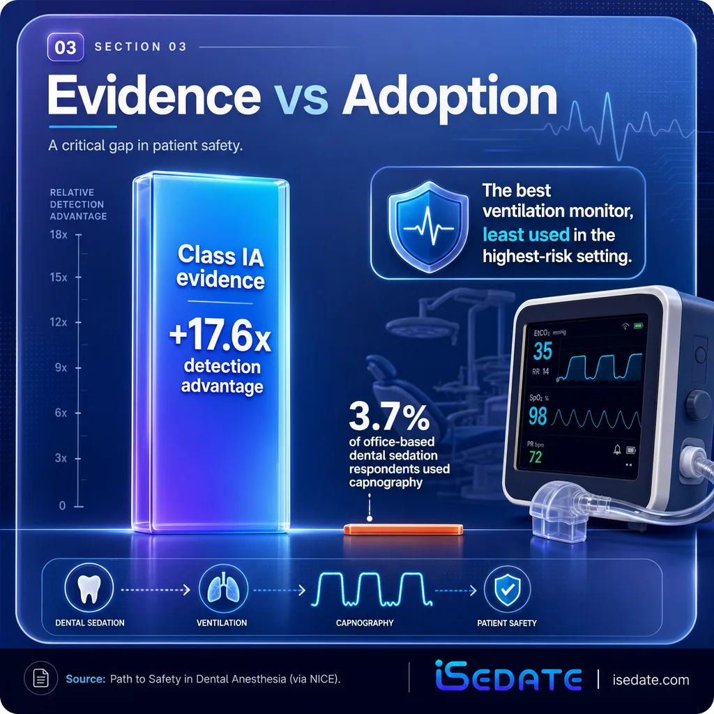
During procedural sedation, respiratory depression is 17.6 times more likely to be detected with capnography than without it, yet one survey found it was used by only 3.7% of office-based dental sedation respondents. That gap between what the evidence supports and what practices actually do is closing fast as requirements tighten. The data below covers how capnography works, what it catches, where it is heading, and its honest limitations.
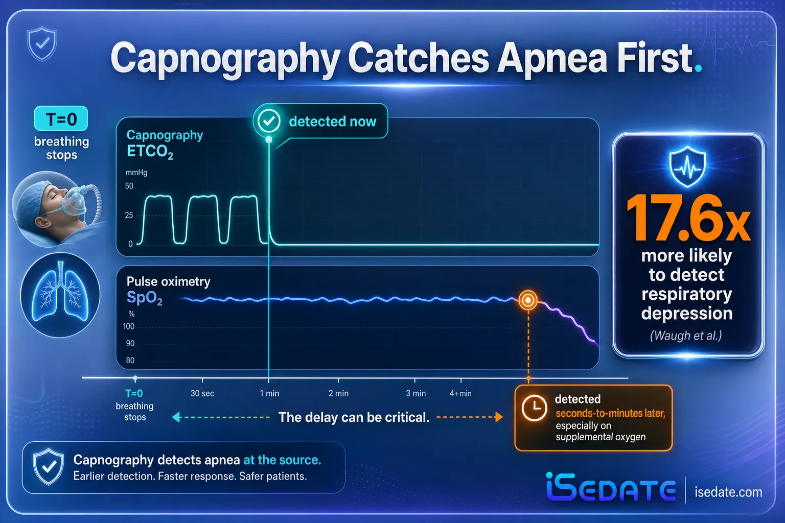
- Respiratory depression is 17.6 times more likely to be detected with capnography than without it during procedural sedation.
- Capnography carries class IA evidence and is described as the standard of care in anesthesia and procedural sedation.
- Historically, capnography was used by only 3.7% of surveyed office-based dental sedation respondents, a large adoption gap.
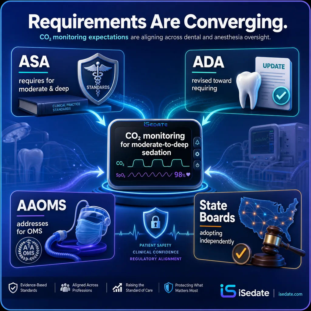
- Requirements are converging: ASA, ADA, AAOMS, and multiple state boards now require or recommend CO2 monitoring for moderate-to-deep sedation.
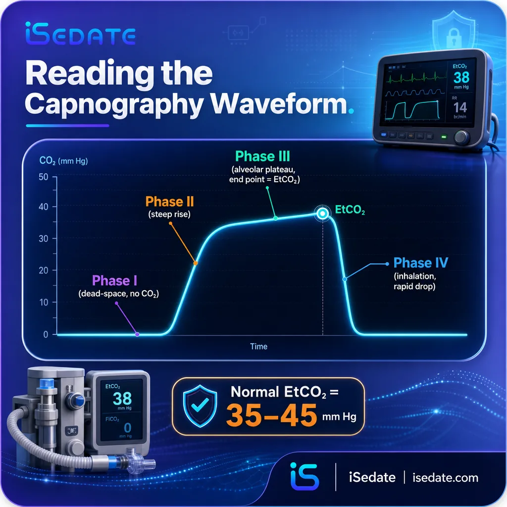
- Normal end-tidal CO2 is 35 to 45 mm Hg; the waveform's four phases let clinicians read ventilation, not just a number.
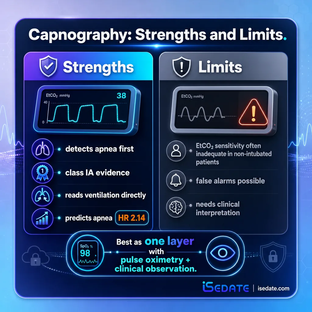
- A hypopnea waveform abnormality raised the risk of subsequent apnea (hazard ratio 2.14), showing capnography's predictive value.
- Honest limitation: in non-intubated sedation patients, EtCO2 can show inadequate sensitivity for some individuals and can generate false alarms, so it complements rather than replaces judgment and pulse oximetry.
What's in This Guide
01 The Detection Advantage
The core case for capnography rests on a single, consistently replicated finding: it catches respiratory trouble earlier and more often than pulse oximetry. The headline number comes from a meta-analysis of procedural sedation studies.
The reason for the advantage is mechanical. Pulse oximetry measures oxygen saturation, a downstream consequence of breathing, while capnography measures exhaled carbon dioxide, a direct product of ventilation. When a patient stops breathing effectively, the capnography waveform changes or ceases immediately, whereas oxygen saturation can lag by tens of seconds to minutes, especially with supplemental oxygen. Multiple randomized trials underpin this, which is why end-tidal capnography is described as having become the standard of care in anesthesia and procedural sedation on the basis of class IA evidence.
Capnography does not replace pulse oximetry, it complements it. Oxygenation and ventilation are different physiologic variables, and a complete monitoring setup tracks both. For the broader framework of which vital signs sedation requires and how continuous monitoring compares to intermittent checks, this piece focuses specifically on the CO2 half of that equation, where the detection advantage is largest.
Source: Capnography in Procedural IV Sedation meta-analysis (PubMed) | End-Tidal Capnography overview (Medscape)
See vitals capture in the anesthesia record
02 How Capnography Reads Breathing
To understand what the numbers mean, it helps to understand what capnography actually shows. It is more than a single value, it is a waveform whose shape tells a clinician about ventilation second by second. Carbon dioxide is a product of cellular metabolism that the lungs eliminate, and the rise and fall of exhaled CO2 traces each breath.
The waveform divides into four phases: exhalation of CO2-free air from anatomical dead space, a steep upward slope as alveolar gas reaches the upper airway, a plateau of CO2-rich alveolar gas whose end point is the end-tidal value, and a rapid drop as inhalation begins. Because EtCO2 is more sensitive to alveolar hypoventilation than oxygen saturation, waveform changes precede desaturation. Research quantified this predictive value: patients showing a hypopnea waveform abnormality, a change of more than 10% from baseline EtCO2, were at significantly increased risk of subsequent apnea, with a hazard ratio of 2.14. The waveform is not just a monitor, it is an early-warning signal.
Treating EtCO2 as a single number misses most of its value. The waveform's shape distinguishes normal breathing from hypopneic hypoventilation, bradypneic hypoventilation, and apnea, each of which carries different risk. In one analysis, waveform patterns during sedation broke down into normal breathing (41%), hypopneic hypoventilation (38%), apnea (15%), and bradypneic hypoventilation (7%). Reading the pattern, not just the value, is what gives capnography its edge.
Source: Capnography and Respiratory Monitoring, StatPearls (NCBI) | Pre-apneic capnography waveform abnormalities (PubMed)
See IV sedation charting with live vitals
03 The Adoption Gap in Dentistry
Given the strength of the evidence, the striking part of the capnography story is how slowly office-based dentistry adopted it. For years, the recommendation and the reality diverged sharply.
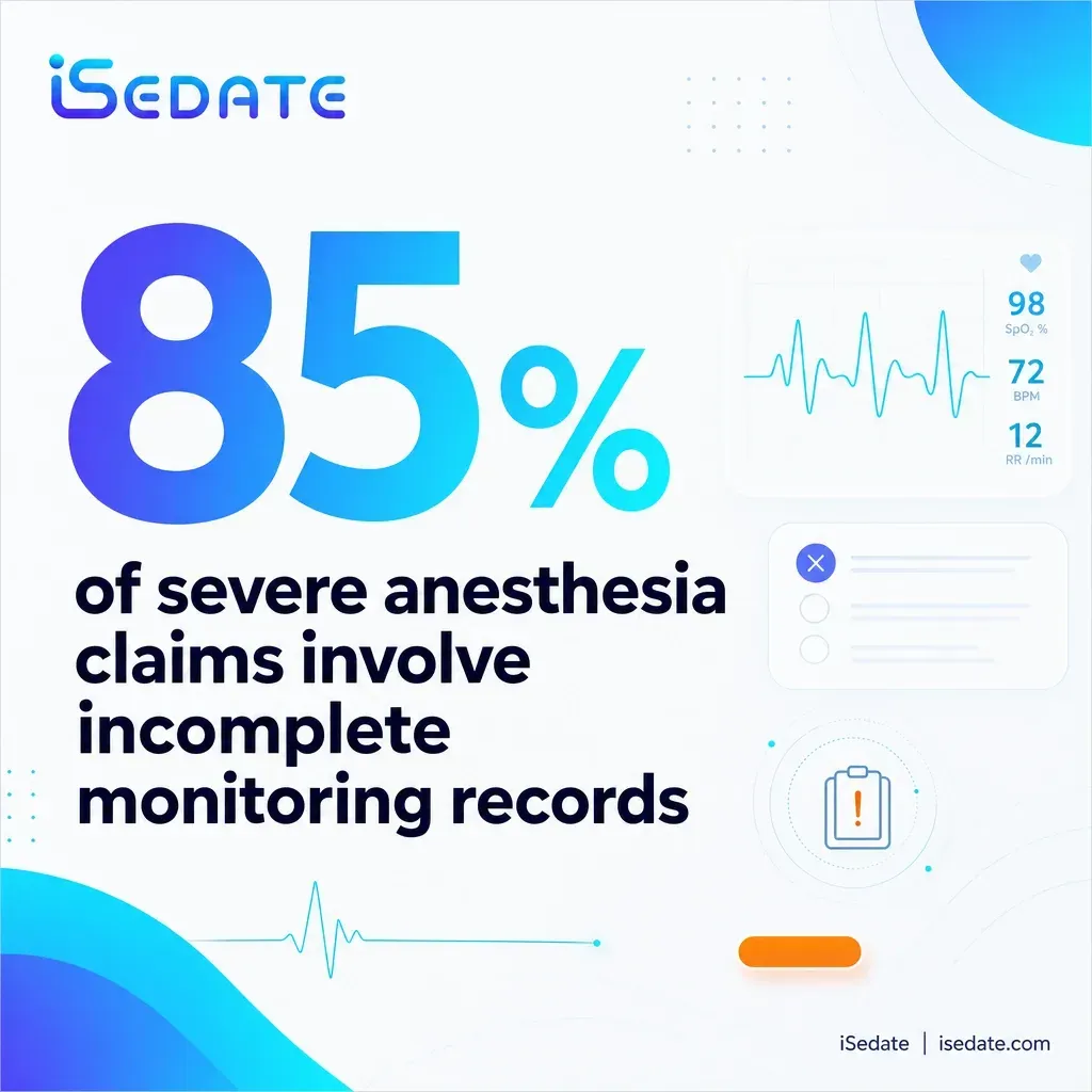
A review of the path to safety in dental anesthesia noted that capnography was used by only 3.7% of respondents in the figure it cited, and characterized standard monitoring in office-based environments as inconsistently utilized. That review directly connected this inconsistency to a demand for stronger policy and guideline statements from professional societies. The gap matters because office-based sedation is precisely the setting where the ASA closed-claims data shows elevated risk, so low adoption of the best ventilation monitor concentrated in the highest-risk environment. The good news is that this figure represents a starting point that requirements are now moving well beyond.
Set two facts side by side. Capnography has class IA evidence and a 17.6x detection advantage, yet a reported 3.7% of office-based dental sedation respondents used it. That is a near-total inversion between evidence strength and adoption, and it existed in the exact setting, the office, where risk is highest. The lesson for a modern practice is that having monitoring capability is not the same as consistently using and recording it, which is the gap standards and documentation systems are now built to close.
Sources: Waugh et al. meta-analysis; The Path to Safety in Dental Anesthesia (Current Anesthesiology Reports). Calculation and interpretation original to iSedate.
Source: The Path to Safety in Dental Anesthesia (Current Anesthesiology Reports)
See how SedationVault records every parameter
04 Where Requirements Are Heading
The adoption gap is closing because the guidance is hardening into requirement. What was once a strong recommendation is increasingly a rule, and the direction across every major body points the same way.
The convergence is broad: the ASA requires capnography for moderate and deep sedation, the ADA moved its guidelines toward requiring CO2 monitoring for moderate-to-deep sedation, AAOMS addresses capnography for oral and maxillofacial surgery practices, and multiple state dental boards have adopted requirements independently. The stated trend is toward universal adoption across all procedural sedation settings. For a provider, this means capnography is shifting from a best-practice option to a compliance expectation, and the practices that adopt it proactively are ahead of the regulatory curve rather than scrambling to catch up.
Cost has been a historical barrier, but the economics have shifted. The EtCO2 monitor is a one-time capital expense, and the recurring cost is disposable sampling cannulas. As requirements broaden, capnography is increasingly treated as a standard operating cost of providing sedation rather than an optional add-on, which reframes the decision from whether to adopt to how to document its use consistently.
Source: AAOMS Capnography Requirements overview | The Path to Safety in Dental Anesthesia
See how oral surgeons use SedationVault
05 The Honest Limitations
A credible case for capnography has to include its limits, because overselling it would be both inaccurate and clinically risky. Capnography is a powerful early-warning tool, not an infallible one, and two limitations deserve attention.
First, sensitivity. A study measuring end-tidal CO2 against directly measured minute ventilation in non-intubated procedural sedation patients found a bimodal distribution, with roughly half of patients showing high instrument sensitivity similar to intubated general anesthesia, and half showing low sensitivity similar to awake volunteers. The authors concluded that in non-intubated patients, EtCO2 often provides inadequate instrument sensitivity to changes in ventilation, meaning the reading does not always track breathing tightly. Second, false alarms. Because the waveform is sensitive, it can flag a transient apnea that never leads to desaturation, and distinguishing alarms that need action from those that do not is described as an essential step for successful implementation.
It cannot. In non-intubated sedation, sampling is less controlled than through an airway device, so EtCO2 can under-represent true ventilation changes for some patients, and false alarms can cause fatigue if every waveform blip is treated as an emergency. Capnography works best as one layer in a monitoring approach that also includes pulse oximetry, clinical observation, and a trained clinician interpreting the waveform, not as a standalone guarantee.
The practical answer to both the adoption gap and the limitations is the same: capture everything continuously, record it, and let a clinician act on the pattern. iSedate's SedationVault pulls live vitals from compatible monitors, including Edan, MindRay, Criticare, and more (the Edan X10 is a common example), so capnography and pulse oximetry data are recorded together as the case proceeds and preserved in a one-click, audit-ready PDF. That continuous record supports real-time recognition and creates the documentation that increasingly required CO2 monitoring will be judged against.
Source: EtCO2 vs minute ventilation sensitivity study (NCBI) | Capnography false alarms during sedation (NCBI)
See SedationVault
06 Summary Table: Every Statistic
| Statistic | Figure | Source | Year |
|---|---|---|---|
| Respiratory depression detection with capnography | 17.6x more likely | Waugh et al. meta-analysis | 2010 |
| Evidence level supporting capnography | Class IA | End-Tidal Capnography overview (Medscape) | 2025 |
| Normal end-tidal CO2 range | 35-45 mm Hg | StatPearls (NCBI) | 2023 |
| Capnography waveform phases | 4 | StatPearls (NCBI) | 2023 |
| Apnea risk with hypopnea waveform abnormality | HR 2.14 | Pre-apneic waveform study (PubMed) | 2019 |
| Waveform patterns: normal breathing | 41% | Cardiac cath lab sequence analysis | 2019 |
| Waveform patterns: hypopneic hypoventilation | 38% | Cardiac cath lab sequence analysis | 2019 |
| Waveform patterns: apnea | 15% | Cardiac cath lab sequence analysis | 2019 |
| Waveform patterns: bradypneic hypoventilation | 7% | Cardiac cath lab sequence analysis | 2019 |
| Office-based dental capnography use | 3.7% | Path to Safety in Dental Anesthesia (via NICE) | 2024 |
| Hypoxia reduction, capnography vs control (endoscopy) | 46% vs 69% | Qadeer et al. | 2009 |
| Desaturation, capnography vs conventional (colonoscopy) | 38.9% vs 53.2% | Beitz et al. | 2012 |
| EtCO2 sensitivity in non-intubated sedation | Bimodal / often inadequate | EtCO2 vs minute ventilation study (NCBI) | 2017 |
| ASA capnography requirement | Moderate & deep sedation | ASA standards / AAOMS overview | 2026 |
| State boards adopting capnography requirements | Multiple, independently | AAOMS requirements overview | 2026 |
07 Frequently Asked Questions
How much better is capnography at detecting respiratory depression?
What is a normal end-tidal CO2 reading?
How widely is capnography used in dental sedation?
Is capnography now required for dental sedation?
Does capnography have limitations during sedation?
All figures trace to primary or peer-reviewed sources. Where a figure originates in a non-dental sedation setting, it is identified as such. Sources include:
- Waugh JB et al. capnography meta-analysis (respiratory depression detection, 95% CI 2.5-122.1)
- "Capnography and Respiratory Monitoring," StatPearls (NCBI)
- "End-Tidal Capnography: Background, Indications, Technical Considerations" (Medscape)
- "Pre-apneic capnography waveform abnormalities during procedural sedation and analgesia" (PubMed, 2019)
- "Sequence analysis of capnography waveform abnormalities" (Scientific Reports, 2019)
- "The Path to Safety in Dental Anesthesia" (Current Anesthesiology Reports, 2024), reporting a capnography utilization figure via NICE
- "The relationship between minute ventilation and end tidal CO2 during procedural sedation" (NCBI)
- AAOMS capnography requirements overview; ASA and ADA monitoring standards
- Qadeer et al. (2009) and Beitz et al. (2012) procedural sedation capnography trials
Capnography's value comes from capturing the waveform continuously and recording it, not just glancing at a number. iSedate's SedationVault pulls live vitals from compatible monitors, builds the record as the case proceeds, and produces audit-ready documentation for every procedure. To see how it works in your practice, book a demo.